
Guide · reading time about 8 minutes
How do I read my spinal MRI?
You are holding your MRI CD, the report is full of technical terms – and you want to understand what is going on. Here we use real images from our practice to show you how to find your way around. And why the image never tells the whole story.

The most important point first
We treat people, not images.
An MRI shows very precisely what your spine looks like. But it does not show which of it is causing your pain. Many changes are part of normal ageing – like grey hair, only on the inside.
That is why the key question is always: does the image match your symptoms and what we find on examination? Only then does a finding become a diagnosis.
Step by step
How to find your way around your MRI
Recognise the plane of the slice
Longitudinal section (sagittal): the spine seen from the side, abdomen on the left, back on the right. Cross-section (axial): like a slice viewed from below. Rarely: Frontal section (coronal), from the front.
Understanding bright and dark
On most images (T2 weighting), water appears bright. That is why the spinal fluid in the canal shines brightly and healthy discs have a bright core. Bone and ligaments appear grey to dark.
Counting vertebrae
Count upwards from the sacrum: the first mobile vertebra above it is L5, then L4 and so on. "L4/5" means the disc between the 4th and 5th lumbar vertebrae.
Mind the sides
In the cross-section you are looking at the body from the feet. Your right side is on the left of the image. A herniation on the "right" therefore appears on the left of the screen.
Compare with your symptoms
A herniation at L5/S1 on the left fits with pain in the left leg down to the calf or sole of the foot. If the image and symptoms do not match, the finding is often of minor importance.
What you see
Your spine on MRI – point by point
On the left the longitudinal section of the lumbar spine, on the right a cross-section. Both images come from our practice.
- Vertebral body – the load-bearing bone.
- Healthy disc – bright, water-rich core.
- Spinal fluid in the spinal canal – bright; the nerve fibres run through it.
- Worn disc – has lost water and looks dark ("black disc").
- Herniated disc – herniated core tissue (nucleus pulposus prolapse) protrudes into the nerve canal.
- Vertebral body/disc in the cross-section.
- Nerve canal – bright triangle with the nerve roots as small dots.
- Facet joint (facet joint).
- Spinous process – you can feel it on your back.
- Deep back muscles (multifidus) – important for stability.
Bright or dark?
Why the same spot looks different
An MRI consists of several image series, called sequences. Each one makes something different visible. The report usually states which were used.
From our practice
Typical findings – and what they look like
In each case the longitudinal section is on the left and the cross-section on the right. The arrows show the decisive spot.
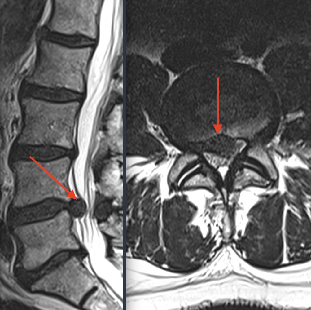
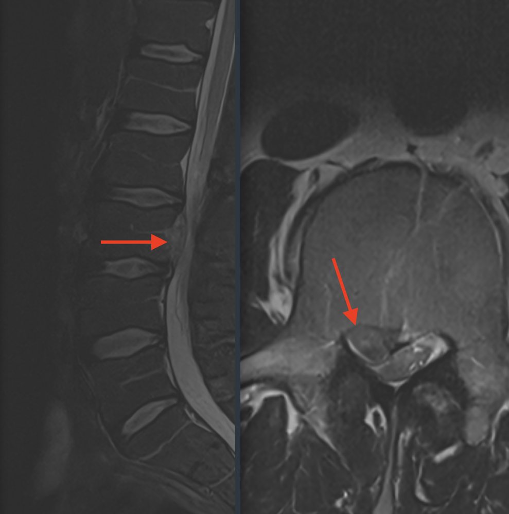
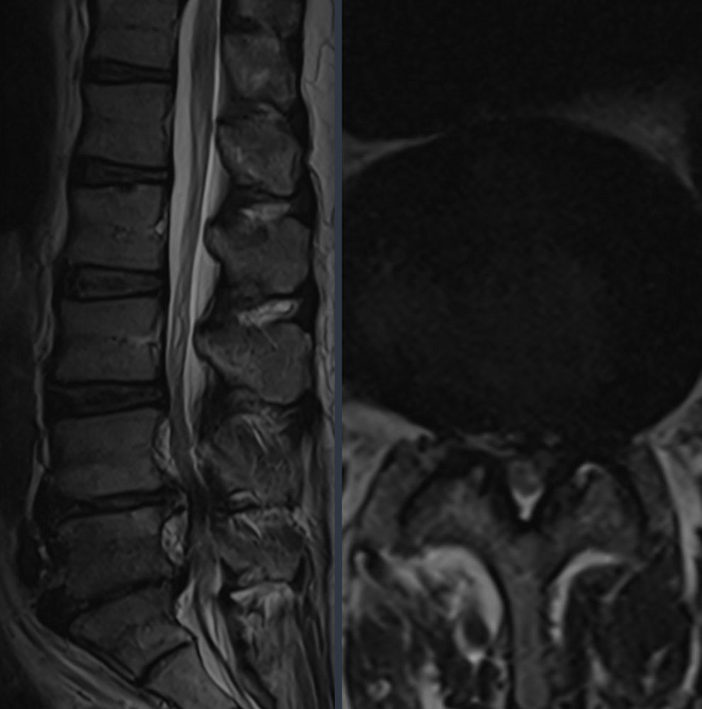
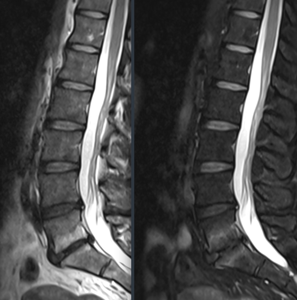
Anonymised images from our practice.
What studies show
Many "findings" are also present in people without back pain
A large review evaluated MRI and CT images of more than 3,000 people without back pain [1]. The result:
What this means for you: a term in the report is no reason to worry as long as it does not match your symptoms. Conversely, a small finding in the right place can cause severe pain. That is why we take time for both – the image and the person.
Report translator
What does this mean in my report?
The most common terms from spinal MRI reports, translated. The coloured label is a rough classification – it does not replace a conversation with your doctor.
- Protrusion · bulge, bulging
- The disc bulges, but the fibrous ring is intact. Very common and often without symptoms. Usually harmless
- Prolapse · extrusion, disc herniation
- Core tissue has escaped through a tear in the fibrous ring and can press on a nerve root. Match with your symptoms
- Sequester · free fragment
- A piece of disc has broken off and migrated in the canal. Often regresses on its own, but can cause strong pressure. Match with your symptoms
- Annular tear · HIZ, annular tear
- Small tear in the outer fibrous ring, visible as a bright dot. A common incidental finding. Usually harmless
- Chondrosis · black disc, disc degeneration
- The disc has lost water and looks darker and flatter. Part of normal ageing. Usually harmless
- Osteochondrosis · Modic changes
- Wear that also affects the adjacent vertebral endplates. Modic type 1 indicates active irritation and can explain back pain. Match with your symptoms
- Facet joint arthritis · facet joint osteoarthritis
- Wear of the small facet joints. Can cause back pain that increases when bending backwards. Match with your symptoms
- Spinal stenosis · narrowing of the nerve canal
- The canal is narrowed by the disc, joints and ligaments. Typical: the legs become heavy or numb when walking, and rest helps. Match with your symptoms
- Lateral recess or foraminal stenosis · lateral canal, neural foramen
- Narrowing where the nerve root exits at the side. Can cause one-sided leg pain. Match with your symptoms
- Ligamentum flavum hypertrophy · thickened yellow ligament
- The ligament at the back of the canal is thickened and often contributes to stenosis. Usually harmless
- Spondylolisthesis · vertebral slippage
- One vertebra has shifted relative to the next. What matters is whether the shift is stable and whether it compresses nerves. Match with your symptoms
- Spondylolysis · defect in the vertebral arch
- A gap in the bone of the vertebral arch, often present since adolescence. Can lead to vertebral slippage. Match with your symptoms
- Synovial cyst · joint cyst
- Fluid-filled outpouching of a facet joint, bright on T2 images. Can press on nerves. Match with your symptoms
- Haemangioma · vertebral body haemangioma
- Benign vascular malformation in the vertebral body, visible as a bright spot. Almost always a harmless incidental finding. Usually harmless
- Schmorl's nodes · endplate fracture
- Disc tissue has pushed into the vertebral endplate. Usually old and of no significance. Usually harmless
- Oedema in the vertebral body · bone marrow oedema
- Fluid in the bone, bright on fat-suppressed series. Can indicate a fresh fracture or inflammation. Have it checked by a doctor soon
- Cauda equina compression · cauda equina compression
- A large herniation or narrowing presses on all nerve fibres in the canal. Together with numbness in the genital area or bladder problems, this is an emergency. Have it checked by a doctor soon
- Myelopathy · cervical spine
- The spinal cord itself has been damaged by narrowing, often visible as a bright spot in the cord. Can affect walking and fine motor skills. Have it checked by a doctor soon
No matching term found. Feel free to ask us at your next appointment.
Well prepared
Five questions for your doctor's appointment
Bring the CD and the written report. We look at the images ourselves – not just the text.
- Which spot on the image explains my symptoms – and which does not?
- Is there anything that needs prompt treatment?
- Which gentle treatments come first?
- How can I tell whether things are getting better or worse?
- Do I need a new scan at all – or only later?
Sources
What this guide is based on
- Brinjikji W et al. Systematic literature review of imaging features of spinal degeneration in asymptomatic populations. AJNR Am J Neuroradiol 2015;36(4):811–816. doi:10.3174/ajnr.A4173
- Chiu CC et al. The probability of spontaneous regression of lumbar herniated disc: a systematic review. Clin Rehabil 2015;29(2):184–195. doi:10.1177/0269215514540919
This guide helps you understand, but does not replace a medical examination and discussion of your findings. As of November 2026.