
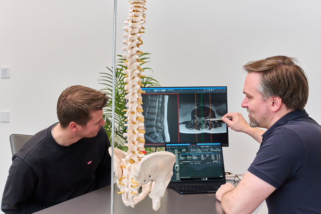
In decompression , a spinal canal that is too narrow is surgically widened. The goal is to give the spinal cord and nerve roots room so that leg pain, numbness and the reduced walking distance subside. Whenever possible we operate endoscopically or microsurgically and preserve the stabilising structures of the spine.
At a glance
- Most common indication: Spinal stenosis of the lumbar spine
- Typical goal: longer walking distance, less leg pain
- Endoscopic or microsurgical through small approaches
- Usually without fusion (spondylodesis)
- Mobilisation on the day of or the day after surgery
What happens in spinal stenosis?
- 1Thickened ligament and joint overgrowth compress the nerve
- 2Widened canal: the nerve has room again
Over the years the yellow ligament (ligamentum flavum) and the facet joints thicken and the disc bulges. Together they narrow the canal. Typical symptoms are pain, heaviness or numbness in the legs when walking and standing, which improve when sitting down or bending forward ("shop window disease of the spine").
When does decompression make sense?
- if walking distance remains limited despite physiotherapy, medication and injections,
- in case of increasing weakness or sensory disturbance in the legs,
- in case of bladder or bowel dysfunction (emergency),
- if imaging and symptoms clearly match.
- 1UnderstandConsultation, examination, imaging, diagnostic injection if needed
- 2StrengthenPhysiotherapy, training, medication, load management
- 3Targeted treatmentInjections, radiofrequency, shockwave, regenerative procedures
- 4Surgeryonly when necessary – as minimally invasive as possible
Our techniques
- Endoscopic decompression: through a working sleeve of about 8 to 10 millimetres, especially for narrowing in the lateral canal or nerve exit foramen.
- Microsurgical decompression: with an operating microscope through a small incision; both sides can often be decompressed through a one-sided approach ("over the top").
- Decompression with stabilisation: only if there is additional instability – see Spinal fusion.
Procedure and aftercare
- PlanningMRI, functional X-rays if needed to check stability, informed consent.
- Surgeryunder general anaesthesia in our partner hospitals in Munich, Arabellaklinik/Herzogpark Klinik and Artemed Klinik München Süd.
- HospitalUp on the day of or the day after surgery, discharge after a few days.
- RehabilitationPhysiotherapy and gait training; follow-up visits at our practice.
Leg pain and walking distance improve markedly in most patients. Back pain due to wear may partly persist – we discuss this openly beforehand.
Frequently asked questions
- How successful is decompression?
- Most patients report a clear improvement in leg pain and a longer walking distance. How good the result is also depends on how long the nerves have already been compressed.
- Does it have to be fused afterwards?
- Usually not. Fusion is only necessary in case of instability.
- How long does recovery take?
- Short walks are allowed immediately. After about six weeks, most everyday activities are possible again without restriction.
Your doctors for this topic



Updated: November 2026 · spectrum MED practice, Sendlinger Straße 37, Munich. This article does not replace a medical examination.